


Slice Computed Tomography (CT)
Overview
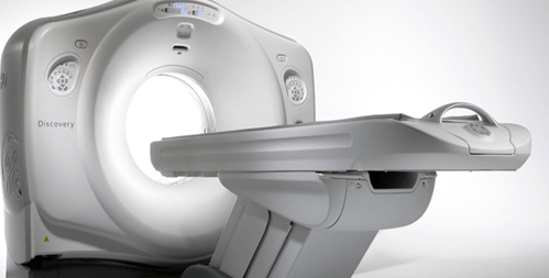
A computed axial tomography scan or CT scan is a medical imaging technology that uses finely focused x-rays together with a detector rather than a photographic film combined with computer imaging technology to create detailed three dimensional images of interior tissue, including soft tissue. It is primarily used to detect and locate structures within the body that cannot be located by other forms of radiological investigations.
Our CT possesses the trademark Gemstone Spectral Imaging technology, which is the essential innovation that drives the high definition breakthrough.
Apart from the advanced imaging capabilities, this technology can be extended across applications and helps in quantitative tissue characterization, anatomical analysis and functional imaging. Importantly, with GSI, patients can have reduced radiation doses and exposure. It comprises a proprietary CT scintillator material. It has an exclusive garnet structure that enables high-definition imaging (0.23 mm) and superfast KV switching. The recovery time is four times as fast as the other detectors and the primary speed is nearly 100 times faster than other detectors. The following are some of the areas where the 128-slice CT is very useful:Oncology:
In oncology, GSI technology helps clinicians in detecting and characterizing lesions, nodules and nodes by improving the visibility of lesions including lymph nodes and hypervascular nodules. Sometimes, by using conventional CT, it becomes quite difficult to distinguish structures whose HU values are too close or very similar – but their composition differs radially. However, GSI technology gives excellent outcomes in such situations – owing to which GSI is useful in studying abdominal anomalies by helping the physicians in getting excellent results.
Nephrology:
GSI is quite helpful in vascular imaging as it optimizes contrast-media utilization – thus enabling exemplary outcomes in precise vascular studies even at low contrast volumes. This is especially helpful in patients with impaired renal function. The usage of high-density material causes errors in measurement studies and the results are not very precise – but GSI employs monochromatic images that helps give precise projection measurements and reduces the errors related to signals.
Cardiology:
There are certain aspects of cardiac imaging that need to be considered for precise cardiac studies to get accurate perfusion and plaque characterization – but, the biggest challenges include calcium blooming and beam hardening. However, GSI overcomes such challenges. It also integrates Snapshot freeze and GSI assist, which make it easy to achieve accurate functional and anatomical stenosis assessment with a single exam.t.