
Patient Information -Diabetic Retinopathy
What is diabetic retinopathy?
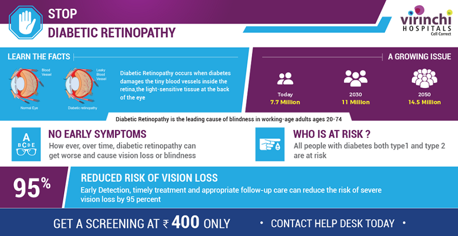
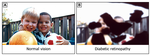
Diabetic retinopathy is an eye problem that can lead to vision loss and even blindness. It affects people with diabetes. It is most common in people who do not control their blood sugar well.
What are the symptoms of diabetic retinopathy?
Most people with diabetic retinopathy have no symptoms until the disease is very advanced. By then, it is usually too late to do anything about the vision loss. That’s why it is important to get screened for the condition early. That way, doctors can take steps to protect your eyes before your vision is damaged.
When symptoms start, they can include:
- Blurry vision
- Dark or floating spots
- Trouble seeing things that are at the center of your focus when reading or driving
- Trouble telling colors apart
When symptoms start, they can include:
- Blurry vision
- Dark or floating spots
- Trouble seeing things that are at the center of your focus when reading or driving
- Trouble telling colors apart
Is there a test for diabetic retinopathy?
Yes. To check for diabetic retinopathy, eye doctors can do 1 of 2 main tests:
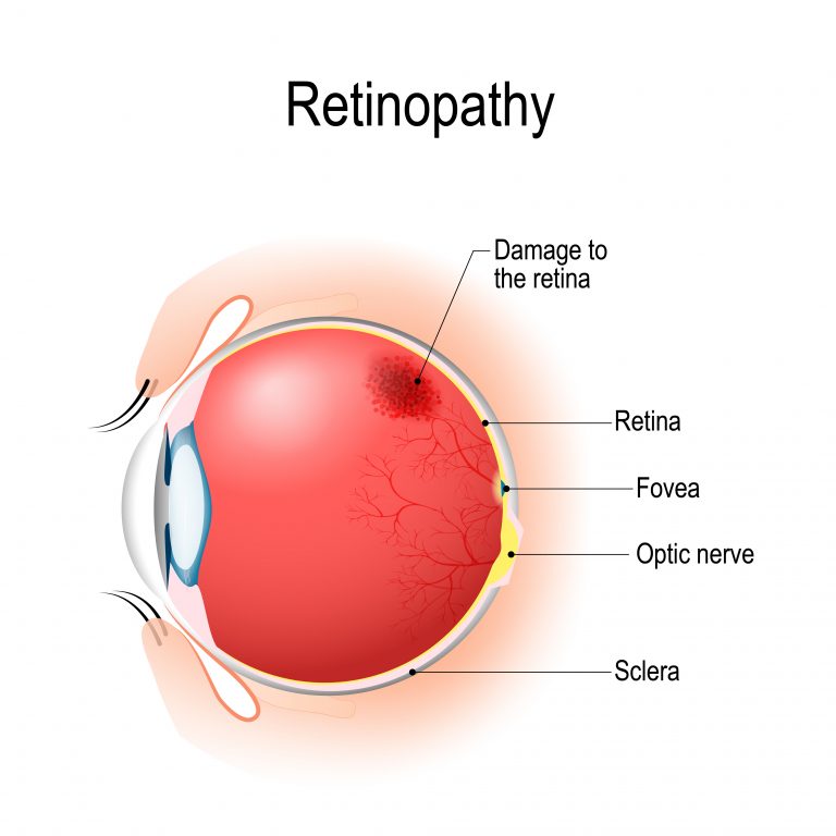
- Dilated eye exam – During this exam, the eye doctor gives you eye drops to make your pupils open up. (The drops make it easier for the doctor to see the different parts of the inside of your eye.) After the drops have done their job, the doctor looks at the back of your eye, called the retina. That’s the part of the eye that is damaged by diabetic retinopathy.
- Digital retinal imaging – For this test, a technician takes pictures of the eye with a special camera. Then he or she sends the pictures to an eye doctor, who checks for disease. It is OK to use this test if your past eye tests have all been normal or if you have no eye doctors nearby. Otherwise, you should have a dilated eye exam.
If either the dilated eye exam or the digital retinal imaging test shows a problem, the eye doctor might suggest other tests, too.
People with diabetes should have their eyes checked regularly. If you have retinopathy, you will need to get checked at least once a year, maybe more. If not, you might only need to get checked every few years. Your doctor will work with you to decide on a schedule. Ideally, checkups should include a dilated eye exam done by an eye doctor. (People who have had normal eye exams in the past and do not have an eye doctor nearby can instead have digital retinal imaging.)
- For people with type 1 diabetes, yearly eye exams should start 3 to 5 years after diagnosis.
- For people with type 2 diabetes, yearly eye exams should start right after diagnosis.
Should I see a doctor or nurse?
If you notice any vision loss (or dark spots in your vision), see an eye doctor as soon as possible.

How is diabetic retinopathy treated?
When mild, diabetic retinopathy is not always treated. But people with the condition do need to keep their blood sugar and blood pressure levels as close to normal as possible. This helps keep the condition from getting worse.
Treatments for diabetic retinopathy can include:
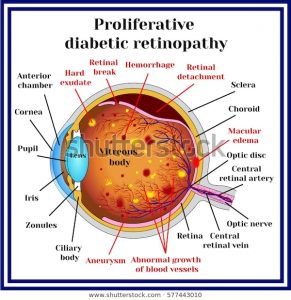
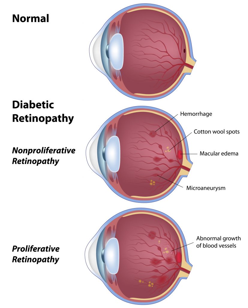
- Photocoagulation – This is laser surgery to seal or destroy leaking or growing blood vessels in the retina.
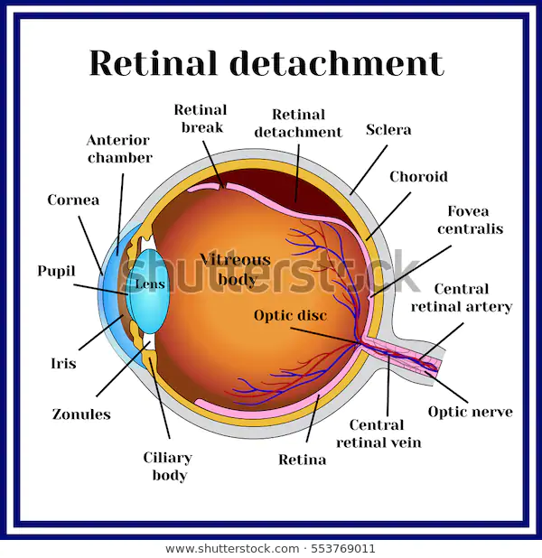
- Vitrectomy – This is surgery to remove blood from the part of the eye called the “vitreous humor” (figure 1). Doctors do this surgery if the blood vessels in the retina leak into the vitreous humor.
- Medicines – Medicines that are injected into the vitreous humor are sometimes used along with photocoagulation or vitrectomy. Your eye doctor will let you know if this type of treatment will help you.
Can diabetic retinopathy be prevented?
Yes. If you have diabetes, you can reduce your chances of getting diabetic retinopathy by keeping your blood sugar and blood pressure levels as close to normal as possible. It might also be important to keep cholesterol levels in the normal range.
Your doctor may do fluorescein angiography or OCT angiography to see what is happening with the blood vessels in your retina. OCT angiography is a newer technique and does not need dye to look at the blood vessels. Fluorescein angiography uses a yellow dye called fluorescein, which is injected into a vein (usually in your arm). The dye travels through your blood vessels. A special camera takes photos of the retina as the dye travels throughout its blood vessels. This shows if any blood vessels are blocked or leaking fluid. It also shows if any abnormal blood vessels are growing.
